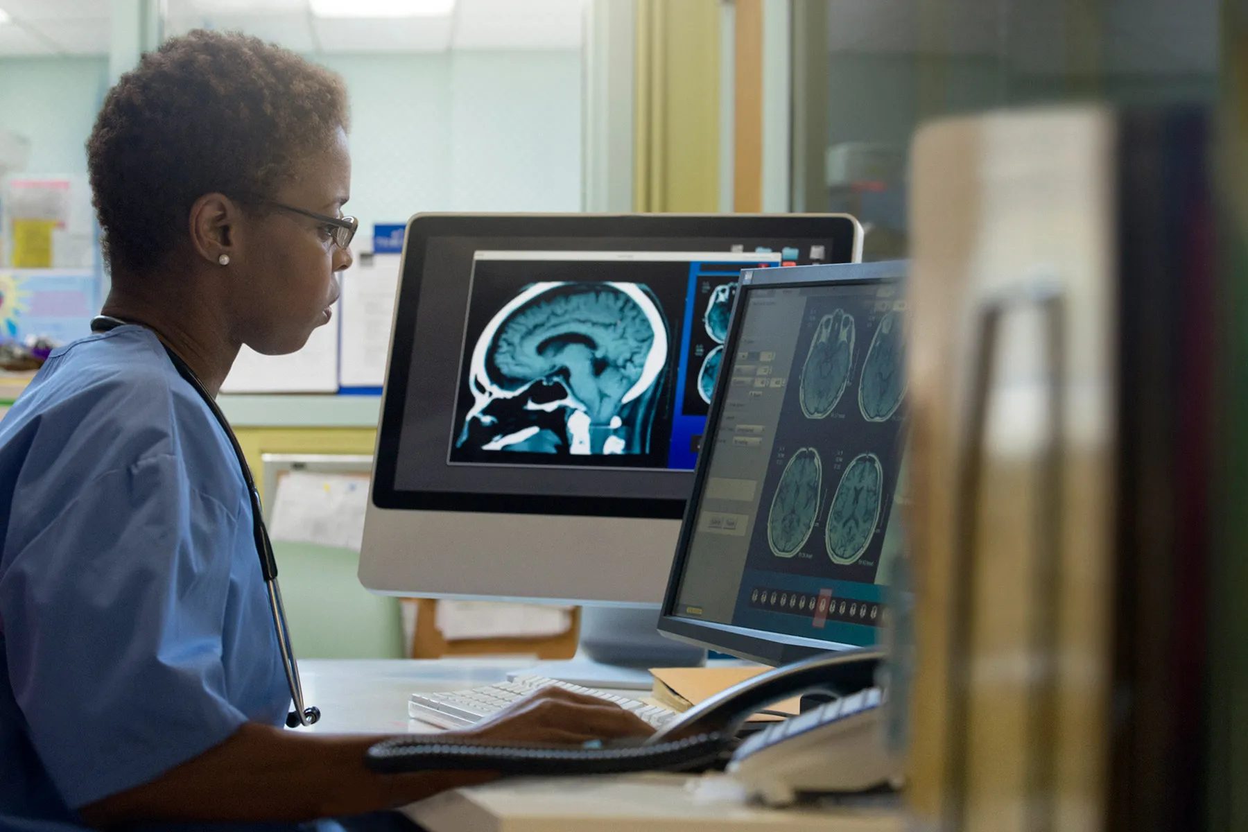
If you have ever been told, “We’re sending you for imaging,” you may have pictured a giant machine, a thin hospital gown, and the vague feeling that medicine has suddenly turned into a science-fiction side quest. Fair enough. But behind that X-ray, CT scan, MRI, ultrasound, mammogram, or PET scan is a specialist whose job is much more important than simply pushing buttons and staring at mysterious gray blobs.
That specialist is the radiologist. Radiologists are physicians who use medical imaging to help diagnose disease, guide treatment, monitor recovery, and in some cases treat problems directly with minimally invasive procedures. In other words, they are part detective, part consultant, part procedural expert, and part translator of your body’s visual receipts.
Many people do not meet their radiologist face-to-face, so the role can seem oddly invisible for someone involved in so many major health decisions. But radiologists are woven into modern medicine in a big way. From confirming a broken bone to spotting a stroke, from evaluating a breast lump to guiding a biopsy, radiologists help answer some of the most important questions in healthcare: What is going on, how serious is it, and what should happen next?
This guide explains what radiologists do, the different types of radiology, why your doctor might send you to one, and what the experience is usually like as a patient.
What Is a Radiologist?
A radiologist is a medical doctor who specializes in obtaining, interpreting, and sometimes using medical images to diagnose and treat disease. That last part matters. A radiologist is not simply “the person in imaging.” This is a physician with extensive training after medical school in imaging technology, anatomy, disease patterns, radiation safety, and clinical decision-making.
Radiologists study how different conditions look on imaging tests. A tiny lung nodule, a hairline fracture, a blood clot, an inflamed appendix, a torn ligament, a blocked artery, or an early breast cancer can each appear in very different ways. Reading those images accurately requires skill, context, and a strong understanding of the patient’s symptoms and medical history.
They also do more than read scans. Radiologists choose or recommend the most appropriate imaging exam, communicate findings to other clinicians, write reports, compare new images with older ones, and sometimes perform image-guided procedures. In many hospitals and clinics, they are the behind-the-scenes specialists helping multiple doctors make better decisions.
The Main Types of Radiologists
Diagnostic Radiologists
Diagnostic radiologists interpret imaging studies such as X-rays, CT scans, MRI scans, ultrasounds, mammograms, and certain nuclear medicine studies. Their job is to identify what is normal, what is abnormal, and what might need more testing or urgent attention. If your doctor orders imaging because of chest pain, abdominal pain, a sports injury, or unexplained symptoms, a diagnostic radiologist is often the one reading the images and sending the report.
Interventional Radiologists
Interventional radiologists are the procedural pros of the field. They use imaging guidance, such as ultrasound, fluoroscopy, CT, or MRI, to perform minimally invasive treatments. Instead of large incisions, they often use needles, catheters, wires, or tiny instruments inserted through a small opening in the skin. These specialists may treat blocked blood vessels, drain abscesses, stop bleeding, perform biopsies, deliver cancer therapies, or help manage conditions such as uterine fibroids. Basically, they are the medical equivalent of people who prefer a keyhole over kicking down the door.
Subspecialty Radiologists
Many radiologists also focus on specific body systems or patient groups. Examples include neuroradiologists, who focus on the brain and spine; breast imagers, who focus on mammography and breast MRI; musculoskeletal radiologists, who evaluate bones, joints, and sports injuries; pediatric radiologists, who specialize in children; and nuclear medicine radiologists, who use radiopharmaceuticals to diagnose and sometimes treat disease. This subspecialization helps match the right expertise to the right problem.
What Radiologists Actually Do All Day
Popular myth says radiologists live in dark rooms, sipping coffee, quietly judging everyone’s insides. The reality is more complex and much more collaborative.
A radiologist may begin the day by reviewing emergency scans for trauma, stroke, or internal bleeding. Then they may consult with surgeons, emergency physicians, internists, oncologists, obstetricians, or pediatricians. They might read a chest CT, compare it with last year’s study, call a clinician about an urgent finding, supervise a fluoroscopy exam, review a mammogram callback, and then perform a biopsy or drain placement in the afternoon.
They are also responsible for image quality and patient safety. That includes helping ensure the right test is used, the right body part is imaged, and radiation is used carefully when a study involves ionizing radiation. They help determine whether contrast material is needed, whether extra views are necessary, and whether a finding deserves watchful follow-up or immediate action.
When you think of radiology as just “picture taking,” you miss the clinical reasoning happening in the background. Radiologists are often connecting the dots between symptoms, lab results, prior studies, and visual clues from inside the body.
Why You Might Need a Radiologist
You usually do not wake up and say, “Today feels like a great day for a consult with radiology.” Usually, another clinician refers you because imaging can answer a question that a physical exam alone cannot.
1. To Find the Cause of Symptoms
If you have severe abdominal pain, persistent headaches, chest symptoms, back pain after injury, or unexplained swelling, imaging can help show what is happening inside the body. A radiologist may help identify appendicitis, kidney stones, pneumonia, disc problems, or a fluid collection that needs treatment.
2. To Confirm or Rule Out an Injury
Broken bones, dislocations, ligament tears, and internal injuries are common reasons for radiology exams. An X-ray may confirm a fracture, while MRI may be better for soft tissue injuries like ligament or tendon damage.
3. For Screening
Radiologists play a major role in screening tests that look for disease before symptoms appear. Mammography is one of the clearest examples. Screening can catch changes early, when treatment may be simpler and outcomes may be better.
4. To Evaluate Cancer
Radiology is central to cancer care. Imaging can help detect a suspicious mass, guide a biopsy, stage cancer, monitor response to treatment, and watch for recurrence. PET scans, CT scans, MRI, mammography, and ultrasound can all be part of the picture depending on the cancer type.
5. To Guide a Procedure
Need a biopsy, drain placement, vascular treatment, or minimally invasive therapy? Interventional radiology may be involved. These procedures can sometimes replace or reduce the need for more invasive surgery.
6. To Monitor a Known Condition
If you have kidney cysts, liver disease, multiple sclerosis, an aneurysm, arthritis, or a treated tumor, follow-up imaging may be ordered to see whether the condition is stable, improving, or changing.
Common Imaging Tests Radiologists Use
X-ray
X-rays are quick and commonly used for bones, the chest, and certain abdominal issues. They use a small amount of ionizing radiation and are often the first-line test when a broken bone, lung problem, or joint issue is suspected.
CT Scan
CT, or computed tomography, uses X-rays and computer processing to create detailed cross-sectional images. CT is especially useful in emergencies because it is fast and excellent for trauma, bleeding, certain infections, kidney stones, lung disease, and many abdominal conditions.
MRI
MRI uses magnets and radio waves rather than ionizing radiation. It is especially good for soft tissues, including the brain, spine, joints, muscles, organs, and some tumors. It can take longer than other studies, and patients may need to answer safety questions about metal implants, pacemakers, or previous surgeries before the scan.
Ultrasound
Ultrasound uses sound waves and does not involve ionizing radiation. It is common in pregnancy, abdominal imaging, pelvic imaging, thyroid exams, blood vessel studies, and many image-guided procedures. It is portable, widely used, and great for real-time imaging.
Mammography
Mammography is specialized breast imaging that uses low-dose X-rays. It is used for both routine screening and diagnostic follow-up when there is a symptom or an abnormality that needs a closer look.
Nuclear Medicine and PET
These tests use small amounts of radioactive material to show how tissues and organs are functioning, not just how they look. That can be especially helpful in cancer imaging, cardiac imaging, and certain neurological evaluations.
Radiologist vs. Radiologic Technologist vs. Radiation Oncologist
These titles sound similar enough to confuse almost anyone who is already wearing non-slip hospital socks, so here is the simple version.
Radiologist: A physician who interprets imaging and may perform image-guided procedures.
Radiologic technologist: The trained professional who performs many imaging exams and helps position you correctly, operate the equipment, and keep the exam running safely and smoothly.
Radiation oncologist: A physician who treats cancer with radiation therapy. That is a separate specialty from diagnostic radiology, though the names are cousins and absolutely love to confuse people.
What Happens Before, During, and After Your Imaging Exam
Before the Exam
Your clinician may explain why the test is being ordered, but the imaging center will usually give you practical instructions. Depending on the exam, you may need to avoid food for a period of time, drink contrast, change into a gown, remove metal objects, or fill out a safety questionnaire. You should tell the team if you are pregnant, might be pregnant, have kidney problems, have a history of contrast reactions, or have implants or devices that could affect MRI safety.
During the Exam
Most imaging tests are painless, though some can be uncomfortable because you need to hold still, lie in one position, or receive contrast material through an IV. CT scans are quick, X-rays are usually very brief, ultrasounds may involve pressure from the probe, and MRIs can feel long and noisy. Interventional radiology procedures are a different category and may involve local anesthesia, sedation, monitoring, and post-procedure recovery.
After the Exam
A radiologist reviews the images and writes a report for the referring clinician. In some settings, patients can also ask questions directly or request to speak with the radiologist. The final report helps guide what happens next, whether that means no further action, repeat imaging, medication, referral, biopsy, surgery, or reassurance that nothing scary is going on.
Questions Worth Asking
If you are heading into a radiology exam, it is completely reasonable to ask:
- Why am I having this test instead of another one?
- Will it use radiation or contrast material?
- Do I need to prepare in any way?
- How long will it take?
- When and how will I get the results?
- Should I mention pregnancy, implants, allergies, or kidney problems?
Good radiology is not only about excellent images. It is also about clear communication, safe care, and using the right test at the right time.
Why Radiologists Matter More Than Most Patients Realize
Modern medicine depends heavily on imaging. A physical exam can tell a doctor a lot, but it cannot directly show a blood clot in the lung, a small brain bleed, a hidden fracture, or the exact location of a tumor. That is where radiologists make a real difference.
They help reduce uncertainty. They help detect disease early. They help avoid unnecessary procedures when imaging is normal. They help guide procedures with smaller incisions and more precision. And they help make sure a clinician is not treating a guess when a clearer answer is available.
In short, radiologists do not just “look at scans.” They help shape diagnosis, treatment, speed, safety, and outcomes across the entire healthcare system.
Experiences Patients Commonly Have With Radiology
To understand why radiologists matter, it helps to look at typical patient experiences rather than just job descriptions. Most people do not remember the official title of the doctor involved. They remember the moment the right test gave an answer.
Imagine someone who slips on wet stairs, lands hard, and swears it is “just a bruise.” The urgent care clinician orders an X-ray. The technologist takes the images, but the radiologist is the one who identifies a subtle fracture that could have been missed by the naked eye. That report changes everything. Instead of walking on an unstable injury for two weeks and making it worse, the patient gets the right brace, the right referral, and the right recovery plan. Not glamorous, but extremely useful if you enjoy keeping your bones in their original arrangement.
Another common experience happens in the emergency department. A patient arrives with severe abdominal pain, nausea, and a very dramatic personal belief that it is either food poisoning or the end of civilization. A CT scan is performed. The radiologist reviews the images and finds appendicitis. That interpretation helps the surgical team move quickly. In another patient, the scan might instead show kidney stones, diverticulitis, or no surgical emergency at all. The point is not that imaging solves every mystery. It is that radiology often narrows the possibilities fast enough to change care on the same day.
Breast imaging is another area where radiologists directly shape patient experience. A screening mammogram may come back normal, offering reassurance. Sometimes it leads to a callback for extra images or ultrasound. That callback can sound terrifying, but it does not automatically mean cancer. Often, the radiologist is being careful and wants a better look at a dense area, cyst, or overlapping tissue. When something truly suspicious is found, the radiologist may guide the next steps, including biopsy and follow-up imaging, helping the broader care team move from uncertainty to a clear diagnosis.
Then there is interventional radiology, which many patients discover only after someone says, “There may be a less invasive option.” A person with a painful abscess may avoid a larger operation because an interventional radiologist can place a drain using imaging guidance. A patient needing a biopsy may have it done with a needle and imaging rather than a more invasive surgical approach. For some people, this means less pain, shorter recovery, and less time in the hospital.
Even routine imaging can matter more than patients expect. A persistent cough leads to a chest X-ray. A sports injury leads to MRI. New neurological symptoms lead to brain imaging. In each case, the radiologist is helping translate pictures into decisions. Patients may never memorize the radiologist’s name, but they often feel the result: a faster answer, a clearer plan, and sometimes major relief.
Final Thoughts
Radiologists are essential medical specialists who help doctors see what cannot be seen from the outside. They interpret imaging, guide treatment decisions, perform minimally invasive procedures, and play a crucial role in everything from emergency care to cancer screening to long-term disease monitoring.
So if your doctor says you need imaging, it is not just a detour on the road to diagnosis. It may be one of the most informative parts of your care. And the radiologist reading that study may be the reason your treatment plan moves from “we’re not sure” to “here’s exactly what we do next.”